Biology homework
Label a cell diagram: identify organelles before you guess
Use shape, position, cell type, and function to label an unfamiliar cell diagram, then check each answer and turn mistakes into practice.

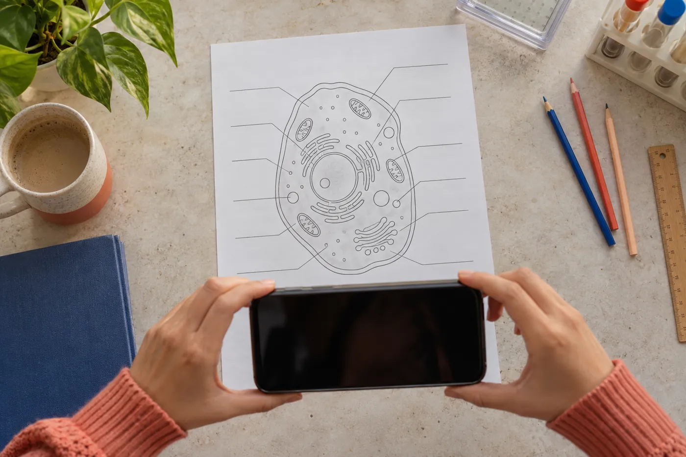
A worksheet shows a large circle, several folded membranes, tiny dots, and ten blank leader lines. You recognize a nucleus, but the endoplasmic reticulum, Golgi apparatus, mitochondria, and small vesicles seem interchangeable. If you guess from color, you may be wrong because textbook artists choose different palettes. To label a cell diagram reliably, use several clues together: cell type, boundary, shape, position, and function.
This guide starts with your own inspection and then shows a careful Lirno workflow when AI help is allowed. Capture the complete task, verify the scan, request one hint or explanation, make your own attempt, and turn the uncertain labels into practice. AI can misread an arrow or oversimplify an organelle, so the worksheet, course vocabulary, and a trusted biology source remain the evidence.
1. To label a cell diagram, capture the whole worksheet
Read the instruction before naming anything. Does it ask for animal-cell structures, plant-cell structures, functions, or a comparison? Look for a word bank, numbered arrows, a key, and any magnified inset. A cropped arrow without the whole cell removes essential context. The same oval could be a mitochondrion, chloroplast, vesicle, or nucleus depending on its internal pattern and the type of cell shown.
If you use Lirno, photograph the entire page in even light with every corner, label number, and instruction visible. Keep the camera parallel so leader lines do not appear to touch a different structure. Review the captured image before asking for help. Correct a blurred number, missing word bank, or cut-off boundary yourself; a complete photo provides context but does not prove the interpretation is right.
State the task in your own words: “I need to label eight structures and give one function for each.” Then make a first pass. Circle labels you know, place question marks beside uncertain arrows, and leave blanks rather than filling them randomly. This separates recognition from uncertainty and gives any requested hint a precise target instead of inviting a generated answer sheet.
2. Decide which cell type the diagram can represent
Before identifying individual organelles, inspect the outer boundary and the large structures. A plant-cell model commonly includes a cell wall outside the plasma membrane, chloroplasts, and a large central vacuole. A typical animal-cell model lacks a cellulose wall and chloroplasts and often has a more flexible outline. Both eukaryotic types contain a nucleus, cytoplasm, ribosomes, mitochondria, endoplasmic reticulum, Golgi apparatus, and a plasma membrane.
Do not classify solely by shape. Artists often draw plant cells as rectangles and animal cells as circles, but real cell shapes vary. Use the presence of several diagnostic structures. OpenStax explains that plant cells have a wall, chloroplasts, and a large central vacuole, while both plant and animal cells share many eukaryotic organelles. That combination is stronger evidence than the outline alone.
Verify what each arrow actually touches. A line ending on the thick outer layer of a plant cell likely targets the cell wall; a second line just inside may target the plasma membrane. If an arrow enters the general fluid region rather than a distinct object, it may indicate cytoplasm. Trace each line from its number to its endpoint before considering the vocabulary list.
Check OpenStax's overview of eukaryotic cells and organelles
3. Match distinctive shapes before the difficult small parts
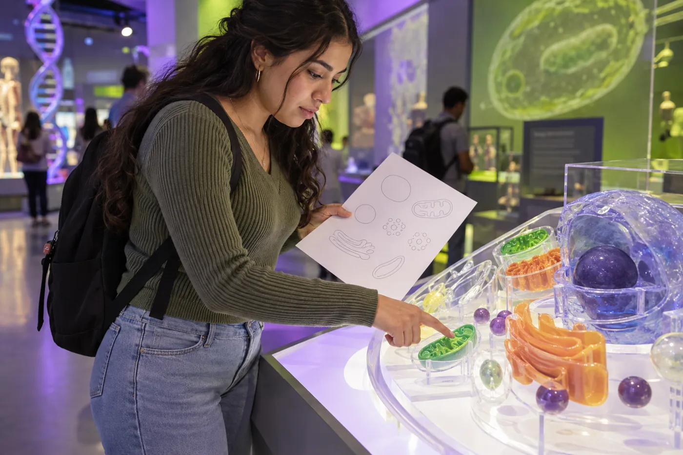
Start with structures that have several clues. The nucleus is usually one of the largest compartments and may contain a darker nucleolus. Mitochondria are often oval with a folded inner membrane. Chloroplasts in plant diagrams may show stacked internal membranes. The Golgi apparatus often appears as a curved stack of flattened sacs with nearby vesicles. These conventions are simplified, but they give you a defensible first set.
Next inspect the endoplasmic reticulum. Rough ER is a membrane network near and continuous with the nuclear envelope, with ribosomes drawn as dots on its surface. Smooth ER lacks those attached dots and is often shown as smoother tubules. Do not label every curved line as Golgi. Ask whether the structure is connected to the nucleus, carries dots, or forms a separate stack.
Leave tiny circles and dots until later. Ribosomes may be free in the cytosol or attached to rough ER; vesicles, lysosomes, and small vacuoles can look similar in a simplified drawing. Use the exact level taught in your class. If the word bank does not include lysosome, do not introduce it merely because a small circle could represent one.
4. Use function as a check, not as a slogan
Attach one accurate function to each provisional label. The nucleus houses DNA and supports control of gene expression. Ribosomes synthesize proteins. Mitochondria carry out stages of cellular respiration and generate much of the cell's ATP. The Golgi modifies, sorts, and packages cellular products. The plasma membrane regulates exchange between the cell and its environment. These statements are more useful than calling every organelle the cell's “control center” or “factory.”
Connect structure to function. Folds in a mitochondrial inner membrane increase the membrane area where key respiration processes occur. Ribosomes on rough ER fit the production of proteins that enter the endomembrane system. A large plant vacuole stores materials and helps maintain pressure against the cell wall. If the proposed function clashes with the visible structure, revisit the label instead of memorizing the mismatch.
Use the wording appropriate to the question. “Makes energy” is too vague because cells transform energy rather than create it. “Produces proteins” may be acceptable for ribosomes, but a longer answer should distinguish synthesis from later modification in ER and Golgi. Your course may expect a simplified function; preserve scientific accuracy without adding details that the task never asks you to assess.
5. Ask for one hint, then make the decision yourself
After your first pass, choose one uncertain arrow. In Lirno, request a clue based on visible features: “What two structures could match arrow 6, and which shape should I inspect?” A hint keeps the identification task with you. An explanation can clarify the difference between rough ER and Golgi. A check can evaluate your completed label list. Select the help mode that fits the stage rather than requesting every answer immediately.
Compare the response with the scan. Did the app follow the correct arrow? Did it confuse a nearby vesicle with the object at the endpoint? If its transcription is wrong, correct the context and reconsider the suggestion. AI may rely on common textbook layouts that differ from your worksheet. The fact that a label sounds plausible is not evidence that it belongs to that particular arrow.
Write your chosen label and a short reason: “mitochondrion—oval with internal folds.” Reasons expose weak guesses. If you can only write “the app said so,” return to the diagram and reference source. Lirno is free to download or start, while some AI usage levels or advanced features may require Premium; neither access level makes an answer automatically correct.

6. Keep plant, animal, and prokaryotic features separate
Build a three-column scratch table if the worksheet compares cell types. All cells have a plasma membrane, cytoplasm, genetic material, and ribosomes. Eukaryotic plant and animal cells have a membrane-bound nucleus and other membrane-bound organelles. Prokaryotic cells do not have a membrane-bound nucleus. This hierarchy prevents the mistaken claim that only animal cells possess a cell membrane or mitochondria.
For the usual school comparison, plant cells have a cellulose wall, chloroplasts in photosynthetic tissues, and a large central vacuole. Animal cells do not have chloroplasts or a cellulose wall. Avoid absolute claims beyond the model: not every plant cell contains visible chloroplasts, and diagrams are teaching representations rather than photographs of every real cell.
Check the vocabulary used in your locale and course. “Cell membrane” and “plasma membrane” may refer to the same boundary; “cytosol” is the fluid component, while “cytoplasm” usually includes cytosol and structures outside the nucleus. If the worksheet distinguishes these terms, follow it. A correct concept placed in the wrong blank can still fail the assignment's intended distinction.
7. Audit every leader line and every paired function
Read the labels in number order and trace each line again. Make sure no two labels accidentally point to the same object unless the diagram intentionally repeats a structure. Check spelling against the word bank and preserve singular or plural where it matters. Then cover the names and see whether each function still identifies the same organelle without relying on color.
Test confusing pairs directly. Rough ER has attached ribosomes; smooth ER does not. The cell wall provides rigid external support, while the plasma membrane selectively regulates passage. Chloroplasts perform photosynthesis, while mitochondria participate in cellular respiration. The nucleolus is a region within the nucleus involved in ribosome assembly; it is not a second nucleus.
If AI checked the page, treat its output as another draft. It can misread a faint arrow, invent a missing label, or state an oversimplified function. Lirno does not guarantee correctness, grades, mastery, or permission to use AI. Follow your school rules and compare final terms with your course material or an authoritative source before submitting.
8. Turn missed labels into retrieval practice
Make one blank card for each label you missed. On the front, sketch the distinguishing clue without writing the name: an oval with inner folds, a stack of curved sacs, or membranes with attached dots. On the back, record organelle, function, and one contrast. Drawing does not need to be artistic; it needs to cue the feature that separates the organelle from its nearest look-alike.
Rebuild the diagram from memory using cards or simple shapes. Place the nucleus, membrane, ER, Golgi, mitochondria, and other required parts, then explain the route of a secreted protein or the difference between a plant and animal cell. This changes the task from recognition to recall. A new worksheet may change colors and orientation, but functions and structural relationships remain useful clues.
After a delay, answer a mixed quiz without the original diagram. You can use Lirno to turn your corrected labels into practice questions, then attempt them before requesting feedback. Include at least one function-to-organelle item, one visual clue, and one cell-type comparison. The goal is not to remember where an artist placed a purple oval; it is to identify structures from evidence on an unfamiliar page.